Cosmetic Non-Surgical Rx Reconstructive Surgical Rx Squamous cell carcinoma Sebaceous cell carcinoma Malignant melanoma Trimalar (tripod) fracture Lacerations |
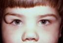
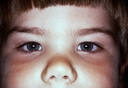
CONGENITAL EYELID AND FACIAL ABNORMALITIESCONGENITAL EYELID COLOBOMA An eyelid coloboma presents at birth as an absence of tissue in either eyelid. In the upper lid these defects are usually full-thickness, while in the lower lid they are characteristically partial-thickness. Defect size may range from a small indentation at the eyelid margin to absence of almost the entire eyelid. This condition occurs most frequently at the inner portion of the upper eyelid or the outer portion of the lower eyelid. They may be associated with other defects of the eye and/or face. Prior to surgery the eye should be protected with lubricant ointment (Lacrilube). Timing of surgery is dependent upon the degree of corneal exposure. The greater the exposure, the more urgent is the surgery. Such surgery involves surgical freshening of the margins of the defect, accompanied by advancement flap reconstruction. The size of this surgical flap is predicated upon the size of the coloboma defect. This surgery is performed under general anesthesia on an outpatient basis. CONGENITAL LACRIMAL OBSTRUCTION Congenital lacarimal obstruction may present as persistent tearing at various times during the first year of life. This condition is caused by delayed or non-opening of the nasolacrimal duct (the valve of Hasner). This is covered under the heading of Lacrimal Reconstruction. CONGENITAL PTOSIS Congenital drooping of the upper eyelid(s) appears at birth and is caused by improper development of the levator muscle. Its surgical correction is covered under the heading of Ptosis. CONGENITAL ENTROPION Congenital entropion is an uncommon condition in which the lower eyelid turns inward resulting in contact between the lid margin and lashes with the cornea. This is caused by improper development of a lower eyelid muscle. It should be distinguished from epiblepharon. While the latter mimics congenital entropion, it is caused by a redundant skin fold at the inner portion of the lower lid whose bulk pushes this part of the lid inward. Both conditions often resolve spontaneously in the first few years of life. Persistent congenital entropion cases can be corrected by the same lid rotation procedure applicable to adult cases. Persistent epiblepharon cases can be corrected by excision of the redundant skin fold. CONGENITAL ECTROPION Congenital ectropion of the lower eyelid is rarely seen at birth as an isolated condition. More commonly it can be linked to congenital eyelid tumors or other congenital eyelid abnormalities including the Kohn-Romano syndrome. Those cases caused by excessive horizontal lid length are surgically treated by horizontal eyelid shortening. Those cases caused by vertical tissue insufficiency are treated utilizing a full-thickness skin graft. Congenital ectropion of the upper eyelid often presents as a total upper lid eversion (outward turning). It often arises as a result of birth trauma. Most cases resolve following conservative measures such as lubricating ointments (Lacrilube), use of a moisture chamber dressing, and temporary use of traction sutures. Persistent cases may require limited surgery. KOHN-ROMANO SYNDROME (BLEPHAROPHIMOSIS SYNDROME) This syndrome is usually hereditary on an autosomal dominant bases. In genetic cases 40-50% of both male and female children may have this condition. It presents at birth with ptosis (drooping upper eyelids), blepharophimosis (horizontal shortening of the eyelids), epicanthus inversus (an abnormal skin fold origination at the inner portion of the lower eyelids that sweeps upward and inward toward the nasal bridge), and telecanthus (widening of the distance between the inner corners of both eyes). Additional more minor manifestations also accompany this disorder. Treatment is surgical - involving tightening the inner corners of both eyes, relaxing the outer corners of both eyes, and ptosis correction. Such surgery affords an excellent functional and cosmetic result. DERMOID CYSTS Dermoids are benign tumors of the orbit that arise during gestational development. Despite this, some appear at birth, while others may present and enlarge many years or decades later. They are caused by skin elements being pinched off as bone segments undergo their normal fusion process. This pinched off tissue continues to grow, albeit in the wrong location. They are most common in the upper-outer portion of the orbit, but can also occur in deeper orbital locations. Surgical excision is the treatment of choice for dermoids. They should be removed in one piece within their own tissue capsule in order to minimize postoperative inflammation. CAPILLARY HEMANGIONA Capillary hemangiiomas are vascular lesions that may be present at birth or appear within the first six months of life. They have a predilictiion for the upper inner orbit, but may also occur in other orbital locations or in eyelid skin or soft tissue. These defects generally present as a bright red elevated area of the skin (strawberry hemangioima). They can also occur on other portions of the face, neck, or back. Some are quite small, while others can be so large that they deform the eyelid and cover the eye. At times this mass enlarges when the baby cries as its vascular channels become engorged. Characteristically, capillary hemangiomas enlarge from 6 months of age to 2 years of age, followed by a period of stagnation and subsequent partial or complete involution (spontaneous diminution in size). Those that do dissipate significantly require no treatment. At times these benign vascular tumors may hemorrhage. This blood accumulation gradually subsides and usually requires no treatment. Yet, treatment is indicated if the hemangioma is large - preventing the child from developing formed vision, causing amblyopia (lazy eye), or significant astigmatism. Treatment is usually centered around steroid injections into the hemangioma (intralesional steroids). Systemic steroids, surgical debulking, and radiation therapy have a negligible role as they provide limited benefit and high risk. |
|
||||